Streptococcus agalactiae
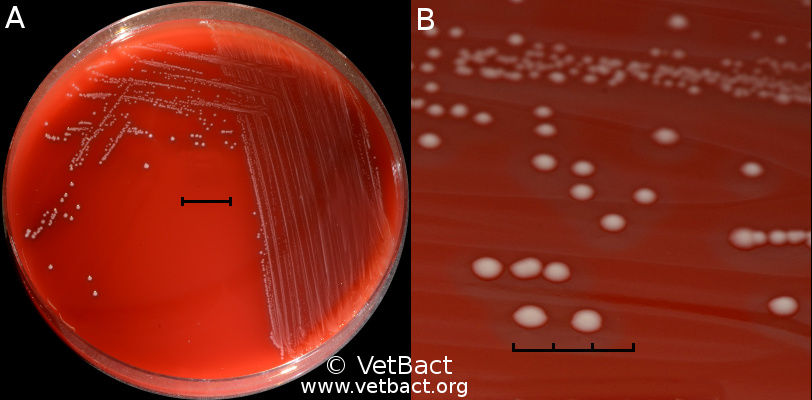
Fig. 16:1. A. Colonies of Streptococcus agalactiae, strain VB 0006/11, cultivated on bovine blood agar at 37 °C during 24 h. The agar plate was photographed with light from above. B. Close-up of some colonies from the agar plate to the left. The β-hemolysis is difficult to see, but it is more easily observed with light from below (see Fig. 16:2). The total length of the scale bars is equivaleent to 1 cm and 3 mm, respectively. Date: 2014-11-14.
Credit: Karl-Erik Johansson (BVF, SLU) & Lise-Lotte Fernström (BVF, SLU).

This work is licensed under a Creative Commons Attribution 2.5 Sweden License.